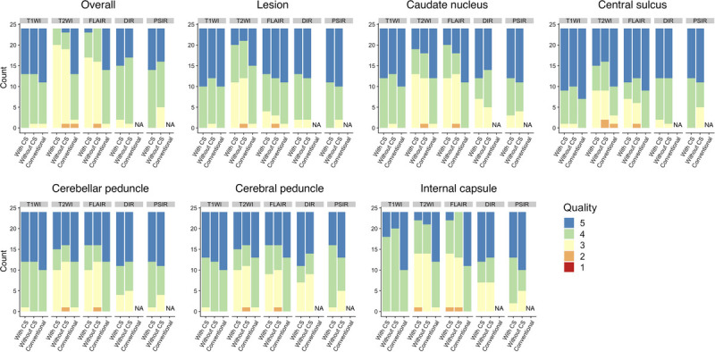
FIGURE 8.

Visual assessment of contrast-weighted images generated from 3D-QALAS with and without CS and conventional imaging for patients with multiple sclerosis. Overall image quality and structural delineation scored on a 5-point Likert score by 2 neuroradiologists are shown. T1WI, T1-weighted images; T2WI, T2-weighted images; FLAIR, fluid-attenuated inversion recovery images; DIR, double-inversion recovery images; and PSIR, phase-sensitive inversion recovery images; NA, not applicable.
