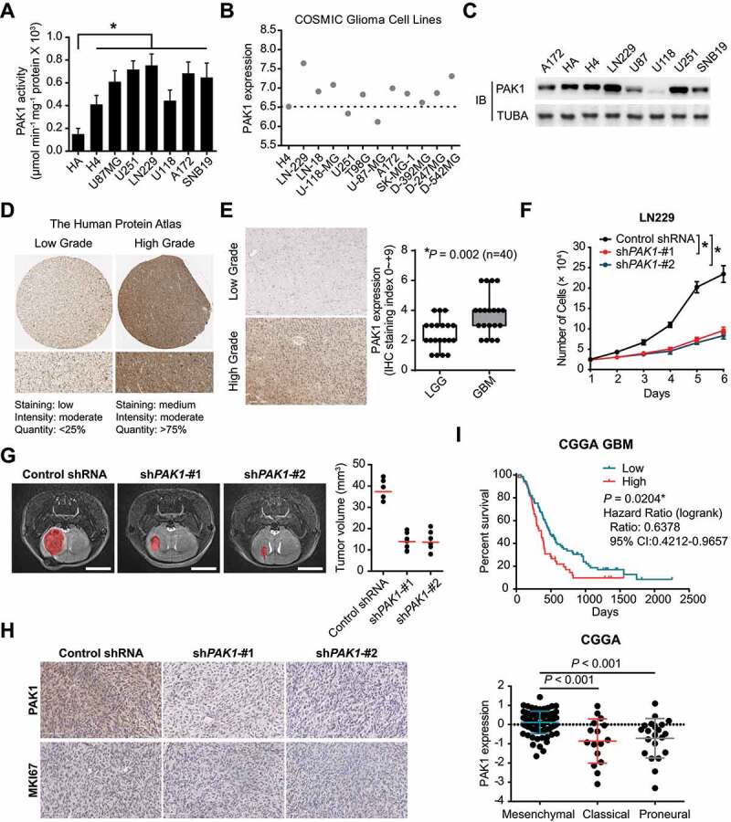
Figure 1.

PAK1 was significantly upregulated and acted as an oncogene in GBM. (A) The kinase activity of PAK1 in GBM cells and normal human astrocytes (HA). (B) The transcriptomic expression of PAK1 in the COSMIC project. (C) The protein level of PAK1 in glioma cells. (D) In the Human Protein Atlas, the representative IHC staining of PAK1 in low-grade and high-grade gliomas was presented. (E) The PAK1 staining in lower grade glioma (WHO II and III) is lower than that in GBM samples. (F) CCK-8 assay indicated that the proliferative potential of LN229 cells was attenuated in PAK1 shRNA groups. (G) The 7 T MR images indicated that the tumor cell growth was inhibited in mice with PAK1 knockdown. Scale bars: 4 mm. (H) The IHC staining of MKI67 in PAK1 shRNA transfected and control tumors in xenograft mice. (I) The prognostic implication and expression pattern of PAK1 in human GBM CGGA data. The OS of GBM patients was shown. The level of PAK1 was higher in the MES subtype compared to Classical or Proneural subtypes
