
Figure 7.
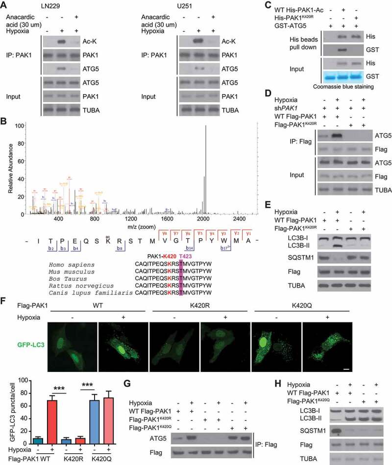
PAK1 at K420 acetylation promotes the binding of PAK1 to ATG5 and autophagy initiation in GBM cells upon hypoxia. (A) LN229 and U251 cells were treated with 30 μM anacardic acid (AA) and subjected to hypoxia or normoxia for one day. Then the indicated antibodies were probed to analyze the cell lysates. (B) MS identified the site of acetylation of PAK1 in hypoxia-treated GBM cells. (C) Ni-NTA agarose beads were used to immobilize bacterially purified WT His-PAK1 or His-PAK1K420R proteins, then incubated with HA-ELP3 and Ac-CoA. Then these beads were incubated with purified GST or GST-ATG5 to perform His bead pull-down assay. (D) The endogenous PAK1 in LN229 cells was knocked down. Then these cells were reintroduced with WT Flag-PAK1 or Flag-PAK1K420R mutant and were subjected to hypoxia or not. Then immunoprecipitation analysis with indicated antibodies was performed. (E) The endogenous PAK1 in LN229 cells was knocked down. Then these cells were reintroduced with WT Flag-PAK1, or Flag-PAK1K420R, and were subjected to hypoxia or not. Western blots were performed as indicated. (F) GFP-LC3 was transiently expressed in PAK1-depleted LN229 cells which were reintroduced with WT Flag-PAK1 or Flag-PAK1K420R or Flag Flag-PAK1K420Q. Representative pictures were presented and quantitation of GFP-LC3 puncta from 10 different images was performed. Scale bar: 0.01 mm. (G) The endogenous PAK1 in LN229 cells was knocked down. Then these cells were reintroduced with WT Flag-PAK1 or Flag-PAK1K420Q mutant were subjected to hypoxia or not. Then immunoprecipitation analysis with indicated antibodies was performed. (H) The endogenous PAK1 in LN229 cells was knocked down. Then these cells were reintroduced with WT Flag-PAK1 or Flag-PAK1K420Q and were subjected to hypoxia or not. Western blots were performed as indicated