
Figure 8.
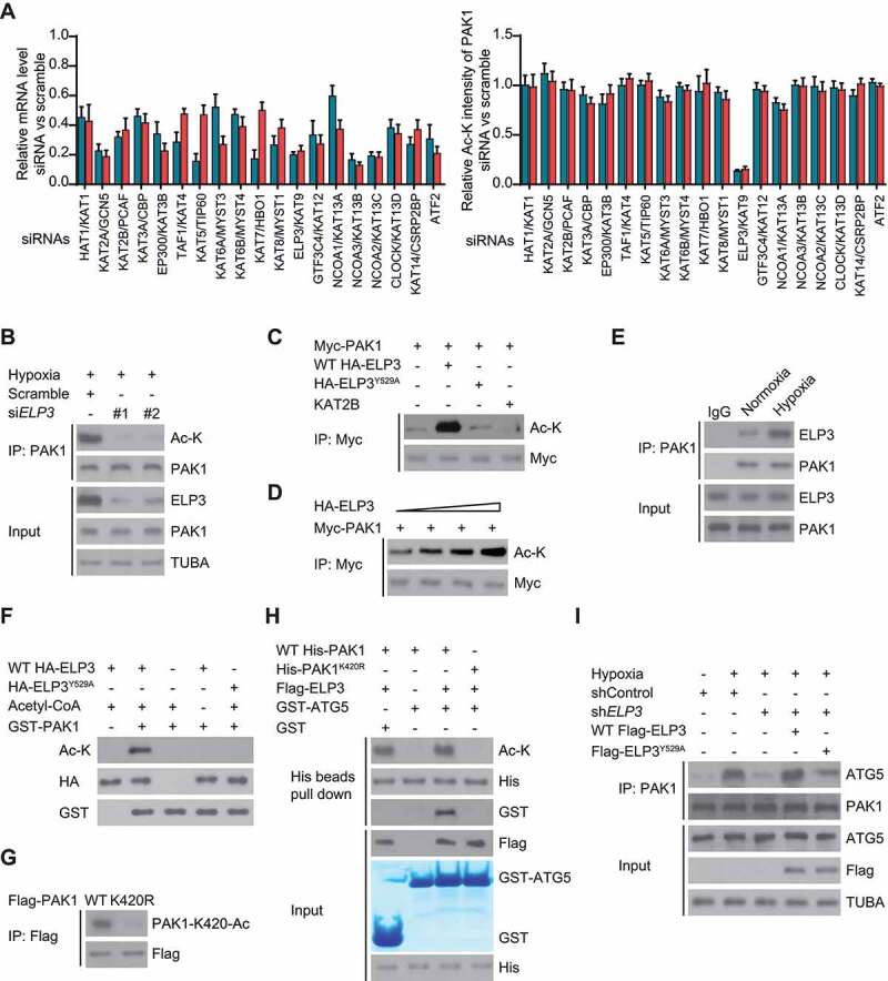
ELP3 is identified as an acetyltransferase of PAK1 in response to hypoxia. (A) One siRNA library with two siRNAs against each of 19 human KAT genes was generated. Each siRNA was transiently transfected into LN229 cells, and the mRNA level of KAT genes was determined by quantitative real-time PCR (left panel). LN229 cells transfected as described were subjected to hypoxia and then harvested for immunoprecipitation with PAK1. Then western blotting was used to detect the acetylation level of PAK1. Band intensity of Ac-K was quantified (right panel). (B) Two different siRNAs targeting ELP3 were transiently transfected into hypoxia-treated LN229 cells. The acetylation level of endogenous PAK1 was determined. (C) ELP3 overexpression increased PAK1 acetylation. Indicated plasmids were transiently co-overexpressed in U87 cells under hypoxia, and PAK1 protein was purified by IP, the acetylation level was determined by western blot. (D) PAK1 with an increasing dose of ELP3 were co-transfected into U87 cells under hypoxia. Acetylation assessment of PAK1 was performed with an Ac-K antibody. (E) Association of endogenous ELP3 with endogenous PAK1 in LN229 cells. PAK1 was immunoprecipitated from LN229 cells treated with or without hypoxia, and the precipitates were analyzed using anti-ELP3. (F) In vitro acetylation assays using purified GST-PAK1 and HA-tagged WT-ELP3 or ELP3Y529A. (G) Flag-tagged PAK1 or PAK1K420R mutant was expressed in U87 cells with ELP3 overexpression. Acetylation assessment was performed with a special PAK1-K420-Ac antibody produced by this group. (H) Ni-NTA agarose beads were used to immobilize purified WT His-PAK1 or His-PAK1K420R. Then these beads were mixed with or without Acetyl-CoA and WT Flag-ELP3, GST or GST-ATG5 to perform the affinity-isolation assay. (I) The endogenous ELP3 in LN229 cells was knocked down. Then these cells were reintroduced with WT Flag-ELP3 or Flag-ELP3Y529A mutant were cultured under hypoxia for 30 min. Immunoprecipitation analysis was performed