
Figure 9.
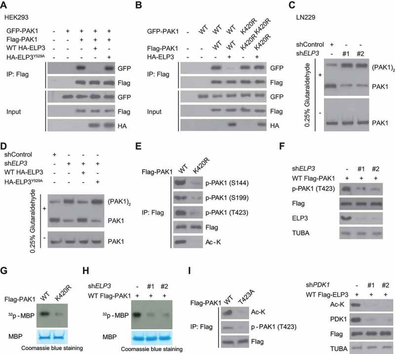
ELP3-mediated K420 acetylation activates PAK1 by decreasing PAK1 dimerization and promoting T423 phosphorylation. (A) WT ELP3, but not its inactive Y529A mutant, suppressed the association of differently tagged PAK1 subunits. WT HA-ELP3 or its catalytic-inactive mutant Y529A was co-expressed with GFP- and Flag-PAK1 in HEK293 cells. Western blot was used to determine the interaction between Flag- and GFP-PAK1. (B) ELP3 co-expression in U87 cells decreased the association of differently tagged WT-PAK1 but not the K420R mutant. Flag- and GFP-tagged PAK1 (WT or K420 R mutant) were co-expressed with or without HA-tagged ELP3. Western blot was used to determine the interaction between Flag- and GFP-PAK1. (C) ELP3 was stably knocked down in LN229 cells using two independent shRNAs. Then these cells were subjected to 0.025% glutaraldehyde treatment. The endogenous PAK1 was immunoprecipitated from LN229 cells, and western blots were used to analyze the levels of monomeric and dimeric PAK1. (D) ELP3 was stably knocked down in LN229 cells. Then these cells were reintroduced with WT-ELP3, or its Y529A mutant and subjected to 0.025% glutaraldehyde treatment. The endogenous PAK1 was immunoprecipitated from LN229 cells, and western blots were used to analyze the levels of monomeric and dimeric PAK1. (E) U87 cells were transfected with WT Flag-PAK1 or PAK1K420R and then subjected to hypoxia. Western blots were performed using indicated antibodies. (F) U87 cells were transfected with ELP3 shRNA and then subjected to hypoxia. Western blots were performed to analyze the T423 phosphorylation of Flag-PAK1. (G and H) In vitro phosphorylation assay. Flag-PAK1 was immunoprecipitated with an anti-Flag antibody from cell extracts in (E and F). Then myelin basic protein (MBP) was used to examine the kinase activity of PAK1 as a phosphorylation substrate. (I) the acetylation levels of K420 of WT Flag-PAK1 or PAK1T423A were determined using immunoblots in hypoxia-treated U87 cells or LN229 cells with or without PDK1 knockdown