Figure 6.

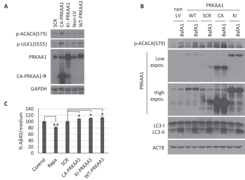
AMPK modulation by gene overexpression in cerebellar granule neurons (CGNs). CGN cultures were infected with lentiviral particles to overexpress the scramble (SCR), constitutive active (CA), kinase-inactive (KI), or WT forms of PRKAA1, and analysis were performed 3 d post-infection. (A) Western blot analysis of PRKAA1 expression levels and AMPK activity with p-ACACA(S79) and p-ULK1(S555). (B) Cells were treated with 10 nM BafA1 for the last 4 h to analyze autophagic flux (LC3-II). Representative western blot of n = 3 independent experiments. Two exposure intensities, low and high, are shown for PRKAA1. (C) Media from APP/PSEN1 CGNs was collected to determine levels of secreted h-Aβ40 by ELISA (n = 3; one-way ANOVA performed; * p ≤ 0.05; ** p ≤ 0.01; Bars represent mean ± SEM)
