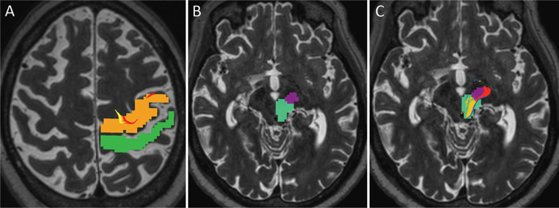
FIG. 5.

Axial T2-weighted MR images showing an example of manually determined ROIs for tractography in an 82-year-old right-handed man with severe bilateral upper-extremity ET. A: ROIs in the precentral gyrus (orange) and postcentral gyrus (green) are depicted on a preprocedure planning image. The DRT (yellow) and pyramidal tract (red) fibers are seen within the precentral gyrus motor area, while the somatosensory fibers tracked posteriorly near the postcentral gyrus but have withered out at this level in this particular patient. Some variability of precise location, size, and extent of tractography-rendered tracts among patients is expected and must be considered on an individual basis. B: Two ROIs were delineated in the midbrain, including the middle portion of the cerebral peduncle for the pyramidal tract (dark purple) and a larger area encompassing the red nucleus and adjacent posterolateral midbrain (turquois) for the DRT and somatosensory fibers. C: Image of the midbrain at the same level with superimposed pyramidal tract (red), DRT (yellow), and somatosensory fibers just posterior to the DRT (blue). Note that the white matter tracts do not encompass the entire ROIs, but rather smaller connecting regions between ROIs. For this reason, manually determined ROIs can span over a greater distance than the expected regions of the tracts.
