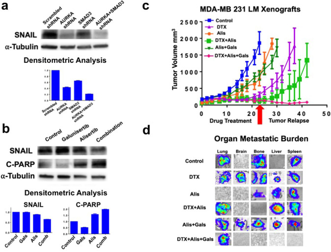
Fig. 6. Dual pharmacologic targeting of TGF-β and AURKA oncogenic pathways.
a Immunoblot assay showing SNAIL protein expression in MDA-MB 231 cells infected with scrambled lenti-shRNAs or lenti-shRNAs targeting SNAI1 for 48 h. Densitometry analysis showing the fold change of SNAIL protein levels in MDA-MB 231 TNBC cells normalized to α-Tubulin. Experiments were performed in triplicate. b Immunoblot assay showing SNAIL and cleaved-PARP expression in MDA-MB 231 cells treated with galunisertib (50 nM) and/or alisertib (50 nM) for 48 h. Densitometry analysis showing the fold change of SNAIL and cleaved-PARP protein levels in MDA-MB 231 TNBC cells normalized to α-Tubulin. Experiments were performed in triplicate. c Establishment of MDA-MB 231 LM xenografts: 1 × 10⁶ cells were injected into the mammary fat pad of 4 weeks old female NSG mice. After 2 weeks of tumor growth, mice were randomized into six groups (five animals each group) and treated with 10 mg/Kg DTX (IP injections), 50 mg/Kg galunisertib (oral gavage), and 50 mg/Kg alisertib (oral gavage) 3 times/week for 3 weeks. After drug treatment, tumor relapse was monitored for additional 3 weeks or when the tumor xenografts reached a volume comparable to control groups. Tumor volume was measured 3 times/week using a digitalized caliper. d Following drug treatment and tumor relapse, mice were sacrificed and organ metastatic burden was determined ex-vivo using the Xenogen imaging system.

