Fig. 7.
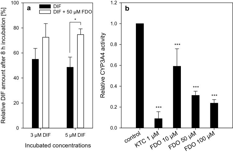
Analysis of toxicokinetic interactions of DIF and FDO. a Quantification of DIF in the cell culture supernatant after 8 h of incubation with HepaRG cells. DIF was incubated alone or in the mixture with 50 µM FDO with HepaRG cells. The measured amount of DIF after incubation with HepaRG cells was normalized to the recovery of DIF from control incubations without cells. Data represent means ± SD (N = 3 independent experiments). Statistics was done by one-way ANOVA with Holm-Sidak post hoc test (all pairwise) with *p ≤ 0.05. b Investigation of CYP3A4 inhibition by FDO. Luciferin-IPA, a specific CYP3A4 substrate, was incubated for 30 min with CYP3A4 supersomes with and without FDO or ketoconazole (KTC). KTC was used as positive control for CYP3A4 inhibition. Treatments with FDO and KTC were normalized to the control without any substance treatment. Statistics was done by one-way ANOVA with Dunnett ‘s test: ***p ≤ 0.001 against control

