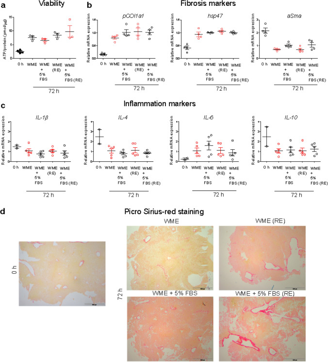
Fig. 1.
Response of tissue slices to different growth conditions. Liver slices were cultured for 72 h in serum-free medium (WME), medium supplemented with 5% FBS (WME + 5% FBS), daily-refreshed serum-free medium (WME RE) or daily-refreshed 5% FBS medium [WME + 5% FBS (RE)] and tested for viability (a), the expression of fibrosis and inflammatory markers (b and c, respectively) and collagen staining (d). a Viability is expressed as the ATP content (pmol) normalized by amount of total protein (µg). The results are the mean and standard error of the mean of 3–5 independent experiments. b and c The expression of selected markers is shown as fold induction over the expression levels in slices cultured in serum-free medium (without refreshing of the medium). The results are the mean and standard error of the mean of 4–5 independent experiments. The relative gene expression was determined by qRT-PCR and calculated using β-actin as housekeeping gene as described in the “Materials and methods” section. Kruskal–Wallis statistic followed by Dunnett’s multiple comparisons test were performed on ΔCt values. d Paraffin sections of liver slices freshly cut (0 h), and maintained in culture for 72 h in serum-free medium (WME) or in medium supplemented with 5% FBS (WME + 5% FBS) non refreshed or daily-refreshed (RE). Red: collagen fibers. Scale bar = 500 µm

