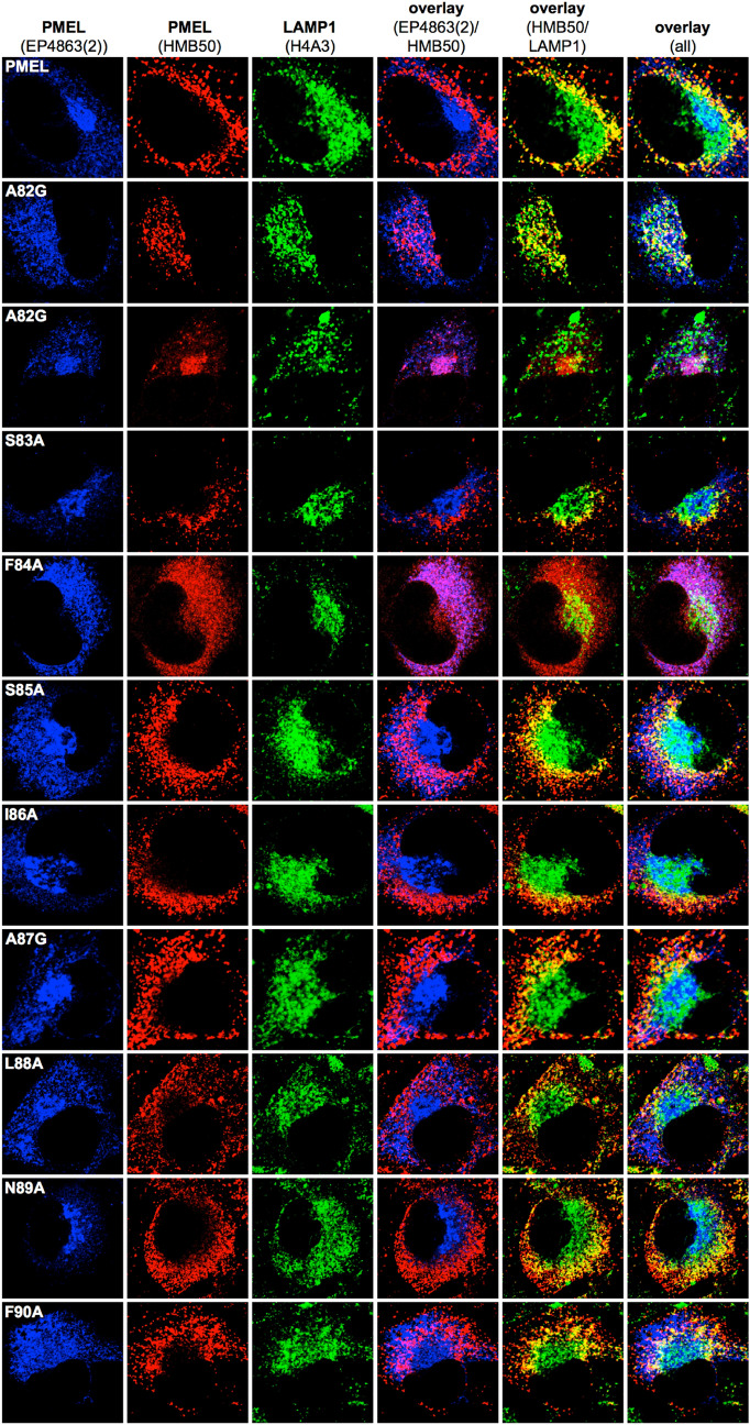
Figure 3.
Subcellular trafficking and melanosome-lysosome segregation of PMEL alanine-scanning mutants. Immunofluorescence analysis of Mel220 cells stably expressing PMEL alanine-scanning mutants. The antibody EP4863(2) recognizes newly synthesized, but not fibrillar PMEL. The PMEL-specific antibody HMB50 recognizes mature melanosomal fibrils. The LAMP1-specific antibody H4A3 recognizes a lysosomal marker. Note that proper PMEL amyloid formation typically correlates in this assay with a significant separation of the perinuclear lysosomal staining (LAMP1) from the largely peripheral melanosomal staining (HMB50).

