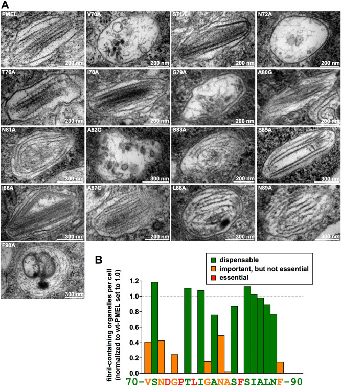
Figure 4.
Melanosomal amyloid formation by PMEL alanine-scanning mutants. (A) EM analysis of Epon-embedded stable Mel220 transfectants expressing PMEL alanine-scanning mutants. Melanosomes of mutants that form fibrils are shown. The respective quantification of fibril formation in the indicated cell lines is depicted in Suppl. Fig. S4 B–E and summarized in (B). (B) Quantitative EM analysis of PMEL alanine-scanning mutants. Shown is the number of fibril-containing organelles per cell [N = 15] after normalization to wt-PMEL (set to 1)). Essential (category 3), relevant (category 2), and largely dispensable residues (category 1) are colored in red, orange, and green, respectively.

