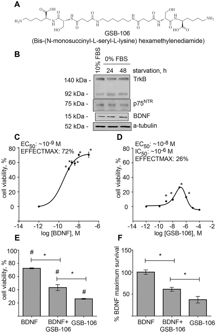
Figure 1.
GSB-106 promotes survival of serum-deprived SH-SY5Y cells. (A) GSB-106 chemical structure. (B) BDNF, TrkB and p75NRT proteins expression in serum starved SH-SY5Y cells. Cells were incubated in serum-free culture medium (“0% FBS”) for 24 and 48 h. After incubation, cells were collected, and protein extracts were subjected to polyacrylamide gel electrophoresis and transferred for Western blotting. Blots were probed with anti-BDNF, anti-TrkB, anti-p75NTR antibodies and anti-a-tubulin antibody. The figure shows data from one independent experiment (n = 3; the original blots are shown in the Supplementary Information file). (C,D) Dose–response survival curves of SH-SY5Y cells (2 × 10⁵/well) treated with BDNF (C) or GSB-106 (D) for 48 h in serum-free condition. Cell viability was measured by MTT metabolism. Cell viability was normalized to viability in control group (“0% FBS”) shown as “10–12 M” (*p < 0.05; n = 7; one-way ANOVA with Newman-Keul’s post-hoc test). (E) Survival of cells (2 × 10⁵/well) incubated with BDNF (100 nmol), GSB-106 alone (100 nmol) and GSB-106 + BDNF (100 nmol for BDNF and GSB-106) in serum-free DMEM for 48 h. Cell viability was normalized to viability in control group (“0% FBS”) (*p < 0.05; #p < 0.05 in relation to control cells (“0% FBS”); n = 7; Wilcoxon t-test). (F) Survival of cells (2 × 10⁵/well) incubated with BDNF (100 nM), GSB-106 alone (100 nmol) and GSB-106 + BDNF (100 nmol for BDNF and GSB-106) in serum-free DMEM for 48 h. Cell viability was normalized to viability in group with BDNF alone (shown as 100% on Y-axis) (*p < 0.05; n = 7; Wilcoxon t-test). Hereinafter (overall) the data is expressed as means ± S.E.M.

