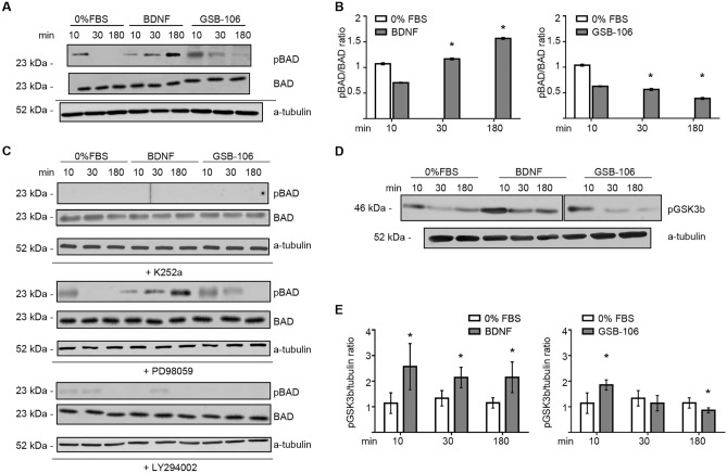
Figure 5.
GSB-106-induced neuroprotection involves the BAD and GSK3β proteins. (A) GSB-106 activates Bad phosphorylation. SH-SY5Y cells were incubated in serum-free culture medium (“0% FBS”) with or without BDNF (~ 1 nmol) or GSB-106 (100 nmol) for 10 min, 30 min, or 180 min. Blots were first probed with anti-phosphorylated BAD (pBAD) antibody and then reprobed with anti-BAD antibody, anti-a-tubulin antibody. The figure shows data from one independent experiment (n = 3; the original blots are shown in the Supplementary Information file). (B) The ratio of signal derived from phosphorylated BAD over BAD bands was calculated (n = 3; *p < 0.05 in relation to “0% FBS”; Wilcoxon t-test). (C) SH-SY5Y cells in serum-free culture medium (“0% FBS”) with or without BDNF (~ 1 nmol) or GSB-106 (100 nmol) were also pre incubated with or without K252a (500 nmol, 60 min), LY294002 (50 µmol, 60 min) or PD98059 (50 µmol, 60 min). The figure shows data from one independent experiment (n = 2; the original blots are shown in the Supplementary Information file). (D) SH-SY5Y cells were incubated in serum-free culture medium (“0% FBS”) with or without BDNF (~ 1 nmol) or GSB-106 (100 nmol) for 10, 30, 180 min. Blots were first probed with anti-pGSK-3β antibody and then were reprobed with anti-a-tubulin antibody. The figure shows data from one independent experiment (n = 3; the original blots are shown in the Supplementary Information file). (E) The ratio of signal derived from p-GSK-3β over a-tubulin bands was calculated (n = 3; *p < 0.05 in relation to corresponding “0% FBS”; Wilcoxon t-test).

