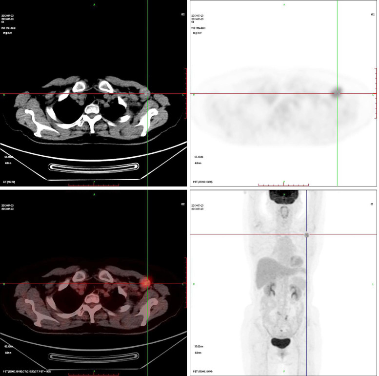
Figure 1.
Positron emission tomography-computed tomography (PET-CT) shows an enlarged lymph node (size, 2.4 cm × 2.4 cm × 2.2 cm) with highly radioactive concentration in the left axilla with irregular shape and uneven density, and the standardized uptake value (SUVmax) is 6.3. No abnormalities are found in the breast.

