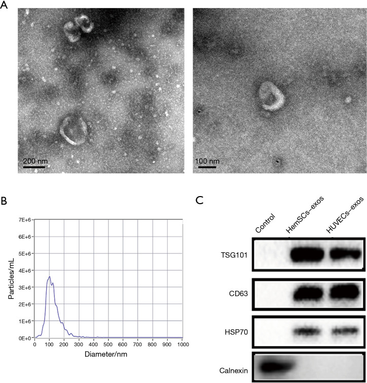
Figure 2.
Identification of exosomes derived from HemSCs. (A) Observation of exosome morphology from HemSCs by TEM (scale bar =200 nm) (left) (scale bar =100 nm) (right). (B) Nanoparticle size analysis displayed that the diameter of the extracted exosomes was around 100 nm. (C) Expression of exosome specific surface markers was determined by Western blot. All experiments were repeated at least 3 times independently.

