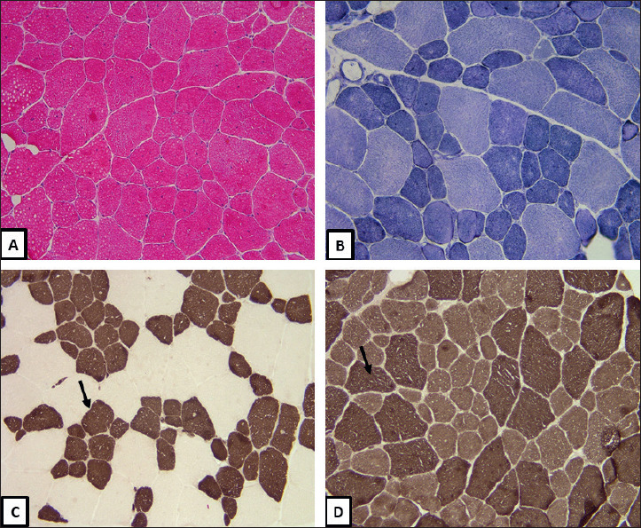
Figure 1.

(A) H&E, Frozen section 200X, show an increase in variation of fiber size and internally and centrally placed nuclei. (B) NADH-TR, Frozen section 200X, ring fibers. (C) ATPase Ph 4.3, Frozen section 200X, type 1 myofibers (arrow) are hypotrophic and more numerous (> 60%). (D) ATPase Ph 9.4, Frozen section 200X type 2 myofibers (arrow) are of normal size and fewer in number.
