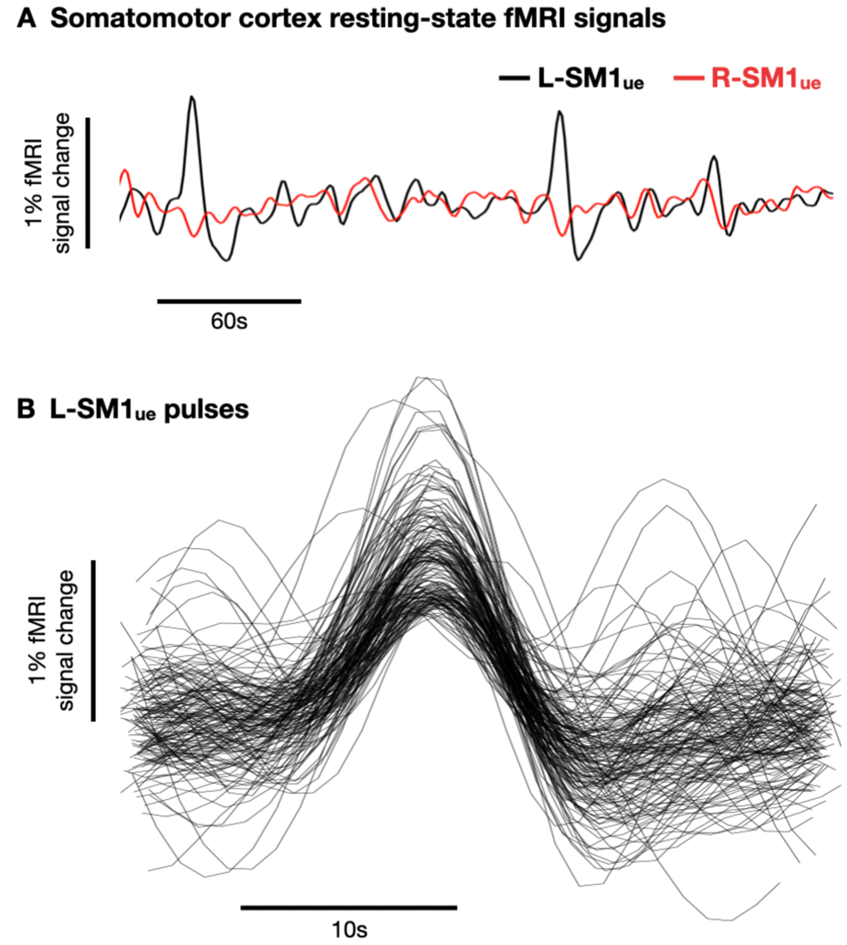
Figure 1. Spontaneous activity pulses in disused somato-motor circuits.

(A) Example resting-state functional MRI (fMRI) signals from left and right primary somatomotor cortex (L-SM1ue and R-SM1ue) during the cast period. (B) Recordings of 144 spontaneous activity pulses detected in an example participant.
