
Figure. Axial Computed Tomographic Images Illustrating the Contralateral Esophageal–Sparing Technique.
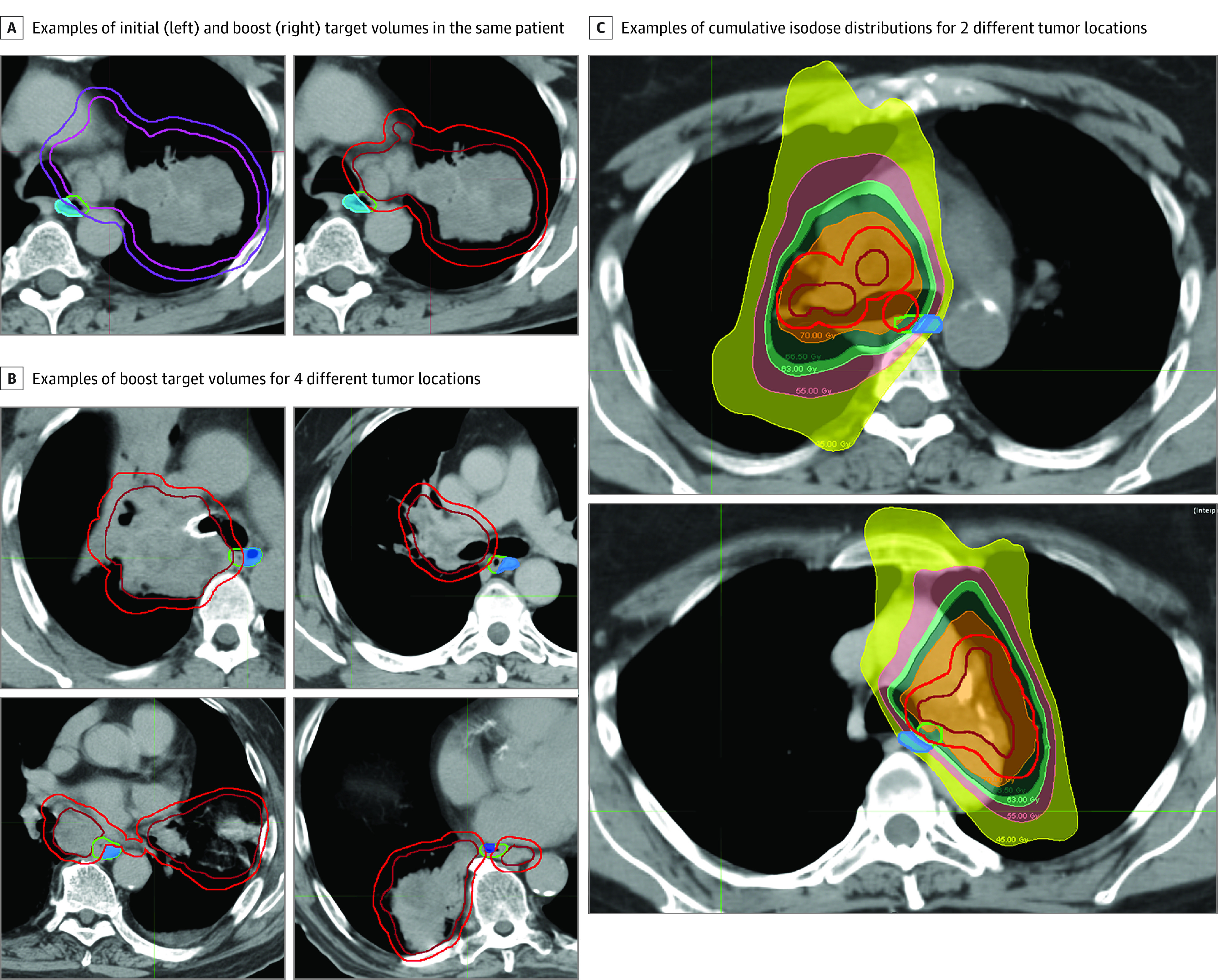
A, Contralateral esophagus (shaded blue), esophagus (green), and target contouring. Left, clinical target volume (pink) and associated planning target volume (purple) treated to 44 Gy. Right, boost internal target volume (red) and associated planning target volume (dark red) treated to 26 Gy. B, Four additional cases with different anatomical tumor locations. C, Two participants with isodose distributions for internal target volume treated to 70 Gy (orange) and planning target volume treated to a minimum of 63 Gy (light green).