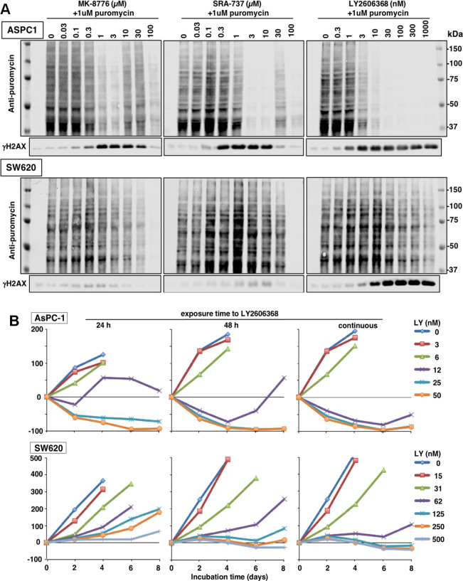
Figure 8.
Inhibition of protein synthesis by CHK1i. (A) ASPC-1 and SW620 cells were incubated with each CHK1i for 24 h. During the final hour, 1 μM puromycin was added to label proteins being synthesized. Cells were analyzed by Western blotting using an antibody to puromycin, followed by fluorescent secondary antibody. Images were generated using a fluorescent scanner. A Coomassie blue stained membrane of the same lysates is shown in Figure S3. (B) AsPC-1 and SW620 cells were incubated with the indicated concentrations of LY2606368 for either 24 h, 48 h, or 8 days. Cells were harvested every 2 days and stained for DNA. A decrease below the starting inoculum reflects cell death.

