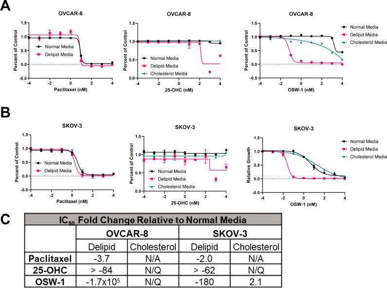
Figure 6.
OSW-1 cytotoxicity is potentiated by the absence of extracellular cholesterol. (A) Cell viability curves of 2D OVCAR-8 cells grown in either normal media (RPMI with 10% FBS), delipidated media (RPMI with 10% delipidated FBS), or cholesterol-supplemented delipidated media (RPMI with 10% delipidated FBS and 20 μg/mL cholesterol), treated with paclitaxel, 25-OHC, or OSW-1 for 72 h. (B) Cell viability curves of 2D SKOV-3 cells grown in either normal media (RPMI with 10% FBS), delipidated media (RPMI with 10% delipidated FBS), or cholesterol-supplemented delipidated media (RPMI with 10% delipidated FBS and 20 μg/mL cholesterol), treated with paclitaxel, 25-OHC, or OSW-1 for 72 h. (C) Fold change compared to cell cultured with normal media (RPMI with 10% FBS) compared to cells treated with paclitaxel, 25-OHC, or OSW-1 in either delipidated media (i.e., Deplid) or exogenously added cholesterol (i.e., Cholesterol). IC50 values used to calculate the fold change are the average of three independent experiments. N/Q = not quantifiable due to curves failing to conform to dose–response curve shape. N/A = not attempted.

