
Fig. 1.
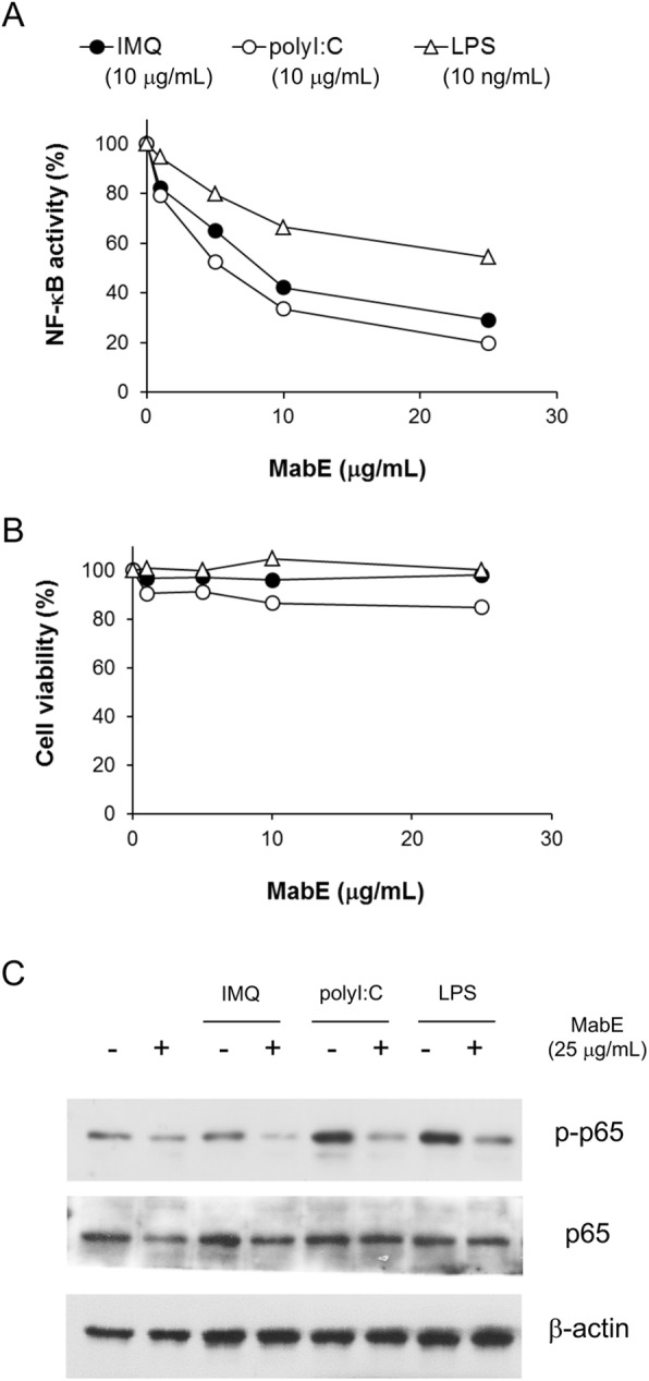
MabE inhibits the TLR ligand-induced activation of NF-κB in RAW264.7 cells. a, b RAW264.7-NFκB-Luc2 cells (5 × 10⁴ cells/well) were seeded onto 96-well plates and pretreated with MabE (1,5,10 or 25 μg/mL). After 1 h, they were stimulated with each TLR ligand (IMQ: 10 μg/mL, polyI:C: 10 μg/mL or LPS: 10 ng/mL). Luciferase activity (6 h) or cell viability (24 h) was measured, and the relative activity or viability compared with untreated control cells was assessed. c RAW264.7 cells (1 × 10⁶ cells/well) were seeded onto 6-well plate and pretreated with MabE (25 μg/mL) or culture medium. After 1 h, they were stimulated with each TLR ligand (IMQ: 20 μg/mL, polyI:C: 20 μg/mL or LPS: 100 ng/mL) for 3 h. Equal amounts of protein in cell lysates were analyzed by Western blotting. The β-actin protein levels were used to confirm that equal amounts of protein were subjected to electrophoresis