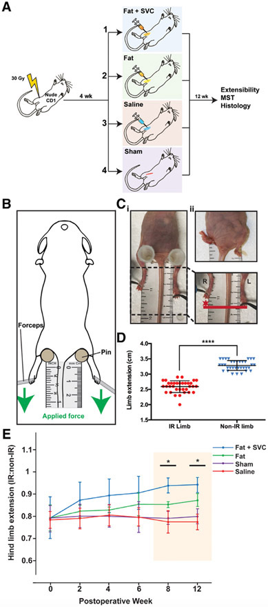
FIGURE 1.
A, The experimental design and four designated animal groups. Total n = 40 (n = 10 mice/group). B, Assessment of hind limb extension. To measure limb extension, mice were stabilized supine using pins to support the hips, with the tail equidistant between the two hind limbs. Equal force was applied to the lower limbs in the caudal direction (green arrow) until resistance was felt, at which point maximum extension was measured (in millimeters) from the middle digit. C, Radiation-induced pronounced hind limb contracture. Representative photographs (i and ii) showing shortening and contracture of the IR (right) hind limb compared with the non-IR (left) hind limb. D, Hind limb extension was significantly reduced on the IR (right) compared with the non-IR (left) side upon assessment of extensibility (****P < .0001). E, Hind limb extension improved over 12 weeks in mice receiving fat grafts. The mice receiving fat + SVCs or fat alone had progressively improved hind limb extension compared with mice in the two control groups. This trend reached significance at weeks 8 and 12 post-grafting (*P < .05). Hind limb extension was not improved beyond baseline in mice receiving saline or sham treatment. IR, irradiated; MST, mechanical strength testing; SVC, stromal vascular cells

