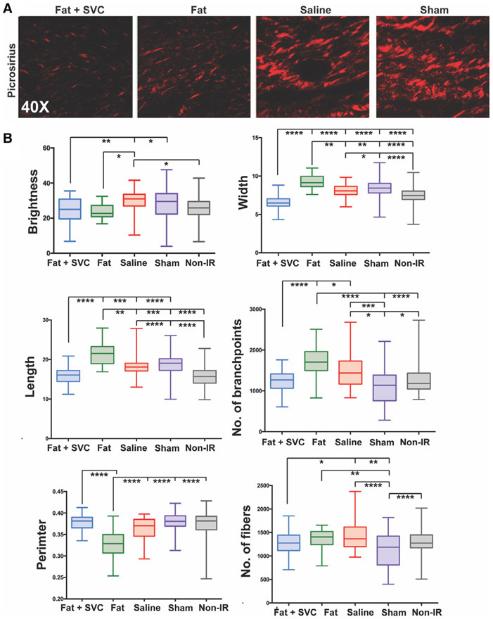
FIGURE 3.
Collagen fiber network analysis. A, Representative images of Picrosirius-stained irradiated hind limb of the mice in the four experimental groups. B, Quantification of collagen fiber network characteristics between the four groups along six parameters: brightness, width, length, number of branchpoints, perimeter, and number of fibers. Irradiated hind limbs grafted with fat + stromal vascular cells (SVCs) had collagen fibers of similar brightness, length, branchpoints, perimeter, and number of fibers to the collagen fibers in non-irradiated mouse hind limb skin, suggesting more regeneration in mice grafted with fat + SVCs compared with control groups of mice receiving saline or undergoing sham treatment. *P < .05, **P < .01, ***P < .001, ****P < .0001

