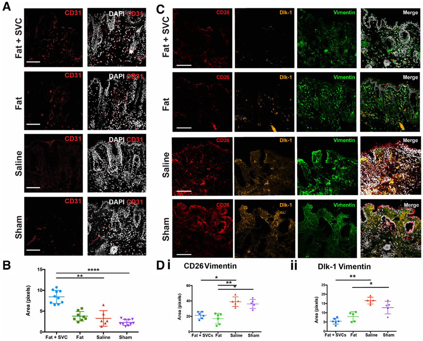
FIGURE 4.
Vascularity of the irradiated hind limb. A, Immunofluorescent staining using CD31 (red) to label endothelial cells and DAPI (white) to label nuclei the four treatment groups. B, Quantification of CD31 staining density revealed significantly increased vascularity in the irradiated hind limb skin of mice grafted with fat + SVCs compared with irradiated hind limbs of mice receiving sham (**P < .01) or saline (****P < .0001) treatment. C, Immunofluorescent staining showing fewer profibrotic CD26+ and Dlk-1+ fibroblast subpopulations in hind limb skin of mice receiving fat grafts. CD26 (red), Dlk-1 (yellow), vimentin (green, to label fibroblasts), and DAPI (white). D, Quantification of the total pixels co-staining for CD26 and vimentin (i) or Dlk-1 and vimentin (ii) between treatment groups. There were significantly fewer CD26+ fibroblasts in the irradiated hind limb skin of mice grafted with fat + SVCs vs mice grafted with saline (*P < .05) and in mice grafted with fat vs mice receiving either saline (**P < .01) or sham (*P < .05) treatment. There were also significantly fewer Dlk-1+ fibroblasts in the irradiated hind limb skin of mice grafted with fat + SVCs vs mice receiving saline (**P < .01) and in mice receiving fat vs sham treatment (*P < .05). Scale bar = 100 μm

