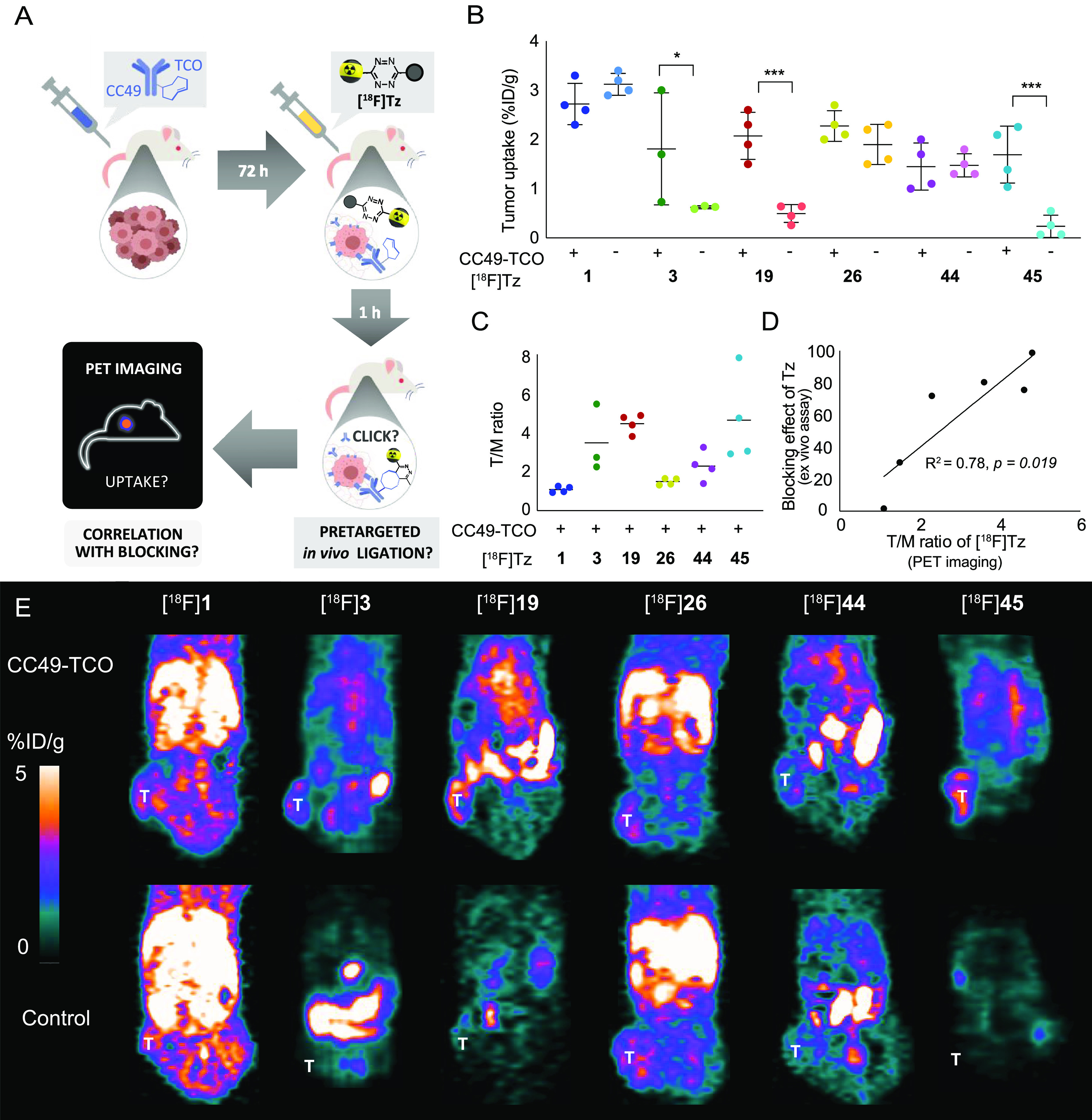
Figure 3.

Pretargeted PET imaging in BALB/c nude mice bearing LS174T tumor xenografts with six 18F-labeled Tz-derivatives. Animals were treated with CC49-TCO or saline (control) 72 h prior to injection of the radiolabeled Tz-derivative. (A) Schematic illustration of the pretargeting experiment and the research question: Is there a correlation between the blocking effect and the PET imaging contras? (B) Image-derived mean uptake values are presented as a percentage of the injected dose per gram (%ID/g) in a tumor 1h p.i. (C) Tumor-to-muscle (T/M) ratios (n = 4 mice, except for [18F]3, n = 3). Data is represented as mean ± SD (*p < 0.05 and ***p < 0.001). For all groups, n = 4 mice (except [18F]3 where n = 3) (D) Correlation between the blocking effect of the unlabeled Tz-derivatives 1, 3, 19, 26, 44, and 45 and the T/M ratios (mean) for the corresponding 18F-labeled compounds observed by in vivo pretargeted PET imaging. A strong correlation was found (linear regression: R2 = 0.78, p = 0.019, n = 6). Data is represented as mean ± SD. The asterisk indicates a significant difference (* p < 0.05 and *** p < 0.001) when compared to control. (E) Representative images of PET scans 1 h p.i. of the radiolabeled Tz ([18F]1, [18F]3, [18F]19, [18F]26, [18F]44, and [18F]45) in pretargeted PET imaging studies (T indicates the position of the tumor). [18F]3, [18F]19, and [18F]45 displayed specific tumor uptake, and the tumor is clearly visualized in the PET image.
