Fig. 3.
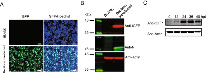
SARS-CoV-2-GFP replicon transfection assay. A Fluorescence microscopy monitoring tGFP expression. 293 T cells were transfected or nontransfected (BLANK) with SARS-CoV-2-GFP replicon, and 36 h post-transfection, cells were fixed, observed and photographed, the nuclei were stained with Hoechst dye, scale bar: 100 µm. B 293 T cells were transfected or nontransfected (BLANK) with SARS-CoV-2-GFP replicon, and 36 h post-transfection, cells were lysed followed by Western blot to monitor the expression of tGFP-BlaR, Nucleocapsid protein (N), and Actin was used as a loading control. C Kinetics of tGFP-BlaR expression, 293 T cells were transfected with SARS-CoV-2-GFP replicon and were lysed at indicated hours post-transfection (hpt) and tGFP-BlaR expression was monitored by Western blot, Actin was used as a loading control.

