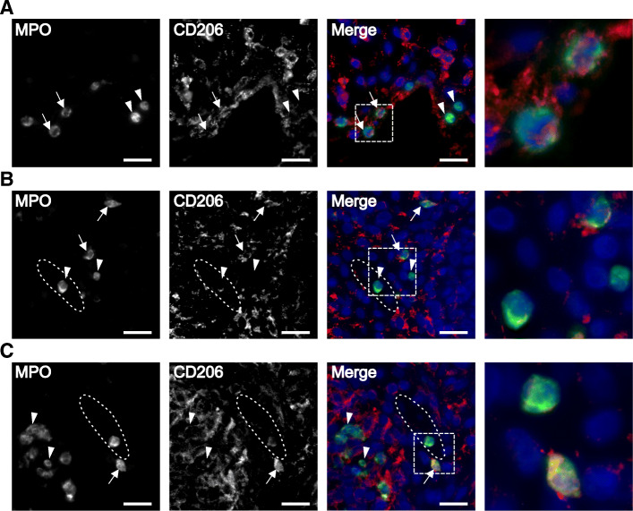
Fig. 2.
CD206-expressing neutrophils are found in synovial tissue. Synovial tissue biopsies from three patients, stained for MPO (green), CD206 (red), and DAPI (blue). Representative images of a patient 4, b patient 8, and c patient 18. Neutrophils expressing CD206, indicated with arrows, and neutrophils without CD206 expression, indicated with arrow heads, were found in all three biopsies. The synovial vessels are indicated with dotted ellipses. Neutrophils were found both scattered in the synovial tissue and inside the synovial blood vessels. The fourth image in each panel represents a magnification of the area indicated in the merged image. Scale bar is 20 μm. All images are taken at × 40 magnification

