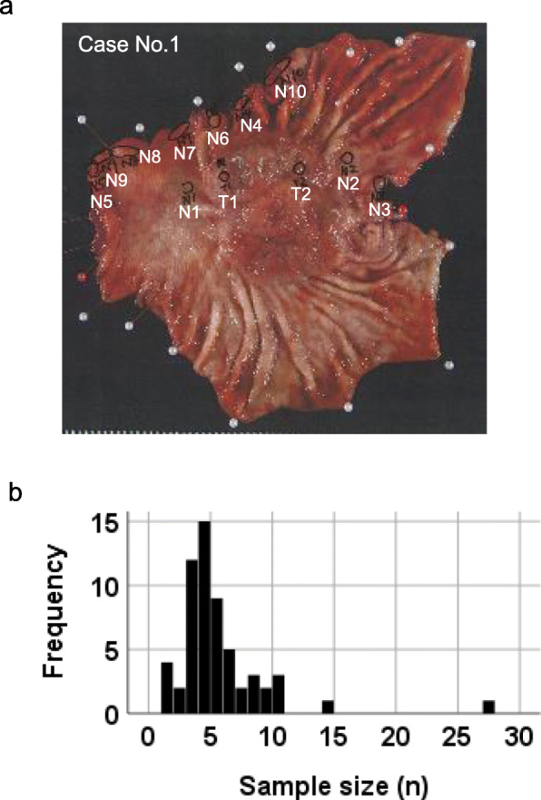
Fig. 1.

Human stomach from which mucosal tissue samples were taken from multiple sites. (a) Example of macroscopic view after multisite sampling in the stomach, resected for gastric cancer in this case. Mucosal tissues were taken: 10 from the nontumor area and 2 from the tumor area (N1-N10, T1-T2). (b) A histogram of the numbers of sampling sites of nontumor areas in gastric cancer cases. The x-axis represents the sample size (numbers taken for DNA analysis) taken from an individual and the y-axis represents the number of individuals with that sample size. Among the 59 cases in this study, the maximum number of samples collected from one case was 27
