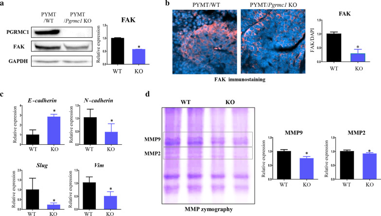
Fig. 3.
Genetic deletion of Pgrmc1 decreases the metastatic markers in the tumors of MMTV-PyMT mice. a Western blot analysis and quantification of PGRMC1 and FAK in the tumors of WT and Pgrmc1 KO mice. GAPDH was used as an internal control. b Immunostaining of FAK in the tumors of WT and Pgrmc1 KO mice. The FAK-positive areas (pink) were quantified. DAPI (blue) was used as an internal control. c mRNA expression of an epithelial marker (E-cadherin) and mesenchymal markers (N-cadherin, Slug, Vim) in the tumors of WT and Pgrmc1 KO mice. Rplp0 was used as an internal control. d Zymographic analysis and quantification of MMP9 and MMP2 in the tumors of WT and Pgrmc1 KO mice. Bars in the right panel show the mean ± standard deviation values (WT, n = 10; KO, n = 16). *, p < 0.05 versus WT mice

