
FIGURE 2.
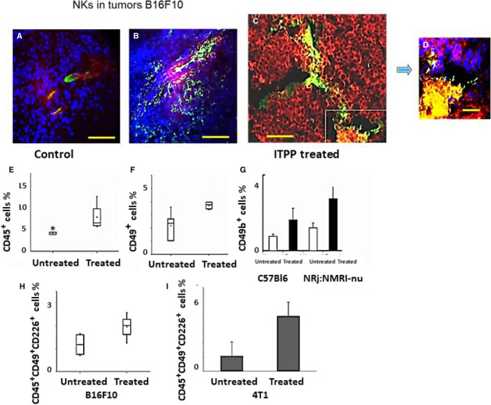
Influence of ITPP treatment of tumours on NK cell recruitment and activation. B16F10 Luc tumours extracted on day 23 were labelled for CD49b+ and CD31+ cell detection (A, B) and for Luc+ expressing cells (C, D). Nuclei were detected by DAPI (A, B, D). Bar = 100 µm (A, B); bar = 40 µm (C); bar = 10 µm (D), pictures are taken with a Zeiss Axiovert fluorescence microscope and analysed by the Zeiss Axiovision software. E, Flow cytometry quantification of the immune CD45+ cells before and after treatment with ITPP. F, CD45+ CD49+ NK cell quantification in B16F10Luc tumours upon ITPP treatment. G, Effect of ITPP treatment on NK cell recruitment in B16F10Luc tumours growing in NRj:NMRI nude mice, compared with normal immunocompetent C57Bl6 mice. H, Activated CD45+ CD49+ CD226+ NK cell quantification in B16F10Luc tumours before and after ITPP treatment. I, In 4T1 mammary carcinoma, activated CD45+ CD49+ CD226+ NK cells were detected by flow cytometry before and after ITPP treatment. Data represent the median values (E, F, H) of five experimental mice from three separate experiments and the means of five experimental mice from three separate experiments (G, I) (*P < .05)