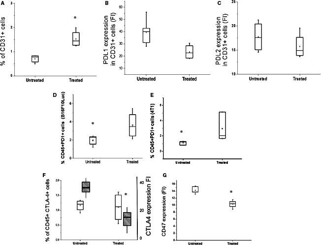
FIGURE 6.

Influence of ITPP treatment on immune checkpoints on distinct cell types in the tumour (A–C) identification of PD‐L1 and PD‐L2 on endothelial cells from a tumour, and modulation by ITPP treatment. Endothelial cells were identified based on their expression of CD31 by flow cytometry (A). The endothelial cell number increased upon ITPP treatment (A). CD31+ endothelial cells expressed less PD‐L1 (B) and tended to express less PD‐L2 (C) after ITPP treatment. Data represent the means of five experiments in triplicate (*P < .05). D, E, The CD45+ immune cell population in the tumour identified by flow cytometry was enriched in PD1‐expressing cells upon ITPP treatment in B16F10 melanoma (D) and 4T1 mammary carcinoma (E). Data represent the median values from five experimental mice and three separate experiments (*P < .05). F, Detection of the CTLA4‐expressing cells among immune cells (CD45+) and level of expression following treatment with ITPP, by flow cytometry. Data represent the means of five experiments in triplicate (*P < .05). G, Detection of the CD47 level expression in B16F10 tumours by flow cytometry. Data represent the median values from five experimental mice and three separate experiments (*P < .05)
