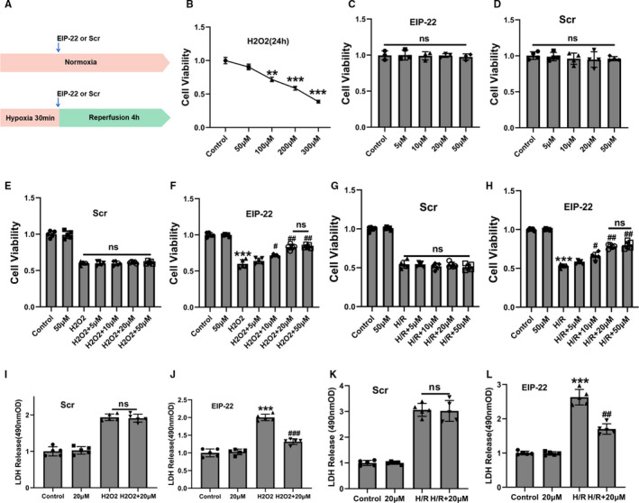
FIGURE 1.

EIP‐22 improved cell viability and reduced LDH release in cardiomyocytes exposed to H/R and H2O2. A, Schematic diagram of H/R model in cardiomyocytes. B, The changes of cell viability in cardiomyocytes were treated with different concentrations of H2O2 for 24 h (50, 100, 200, 300 μM). C, Effects of different concentrations of EIP‐22 on cardiomyocytes viability (5, 10, 20, 50 μM). D, Effects of different concentrations of Scramble peptide (Scr) on cardiomyocytes viability (5, 10, 20, 50 μM). E, Effects of different concentrations of Scr on cell viability in H2O2 model. F, Effects of different concentrations of EIP‐22 on cell viability in H2O2 model. G, Effects of different concentrations of Scr on cell viability in H/R model. H, Effects of different concentrations of EIP‐22 on cell viability in H/R model. I, Effects of 20 μM Scr on LDH release in H2O2 model. J, Effects of 20 μM EIP‐22 on LDH release in H2O2 model. K, Effects of 20 μM Scr on LDH release in H/R model. L, Effects of 20 μM EIP‐22 on LDH release in H/R model. The data represent means ± SD. ** P < .01 vs. the control group, *** P < .001 vs. the control group, ## P < .01 vs. the H2O2 or H/R group, ### P < .001 vs. the H2O2 or H/R group, ns, not statistically significant
