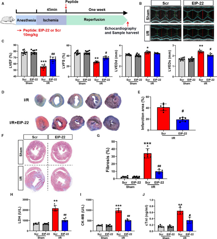
FIGURE 4.

EIP‐22 ameliorates myocardial I/R injury. A, Myocardial I/R model diagram. LAD for 45 min, reperfusion for 1 wk, the concentration of EIP‐22 peptide is 10 mg/kg. B, The representative photographs of echocardiography. C, Quantification data of echocardiography analysis. D, The representative photographs of Evans Blue‐TTC staining. E, Quantification data of TTC staining. F, The representative photographs of Masson staining. G, Quantification data of Masson staining. H, The detection of serum LDH level. I, The detection of serum CK‐MB level. J, The detection of serum cTnI level. The data represent means ± SD. * P < .05 vs. the Sham + Scr group, ** P < .01 vs. the Sham + Scr group, *** P < .001 vs. the Sham + Scr group, # P < .05 vs. the I/R + Scr group, ## P < .01 vs. the I/R + Scr group
