
FIGURE 6.
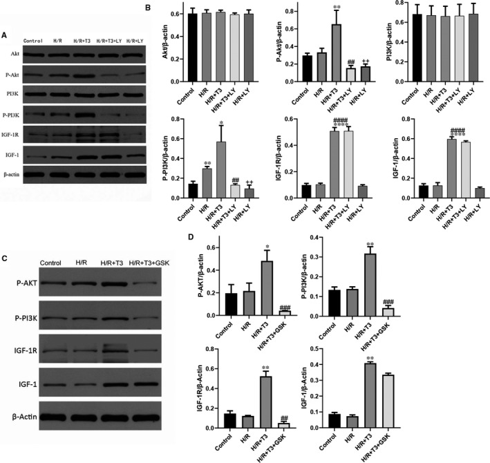
T3 protected cardiomyocytes from H/R‐induced apoptosis through PI3K/Akt signalling pathway (A and B). A, The expression of Akt, P‐Akt, PI3K, P‐Akt, IGF‐1, IGF‐1R; β‐actin protein expression was used for normalization and (B) representative bands quantified in the corresponding bar graph. The T3‐induced activation of PI3K/Akt signalling was mediated by IGF‐1/IGF‐1R (C and D). The expression of P‐Akt, P‐PI3K, IGF‐1 and IGF‐1R was analysed by Western blotting; and representative bands quantified in the corresponding bar graph. β‐actin protein expression was used for normalization. The values were expressed as the mean ± SD in the experiments. n = 6. ··P <.01 vs control; * P <.05, ** P <.01, **** P <.0001 vs H/R; ## P <.01, ### P <.001 vs H/R + T3