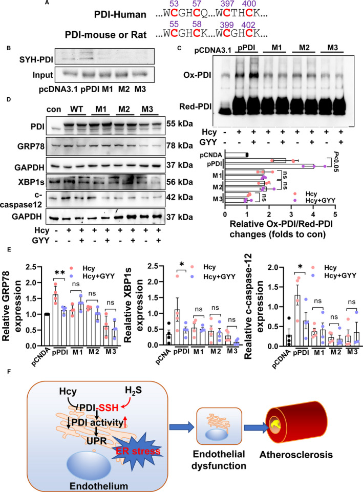
FIGURE 6.

PDI sulfhydration sites and their activity. A, Two conserved cysteine‐terminal CXXC in humans, rat and mouse. B, H2S induced PDI sulfhydration changes by transfection wild‐type human PDI (pPDI) plasmid, mutation C53/57 (M1) plasmid, mutation 397/400 plasmid (M2) and mutation four cysteine site (M3) plasmid into HEK‐293 cells. C, H2S‐promoted PDI activity changes while mutation sulfhydration sites of PDI. D, H2S reduced HHcy‐induced ER stress while mutation sulfhydration sites of PDI. E, The relative protein expression of GRP78, XBP1s and cleaved‐caspase‐12. **P < 0.01, *P < 0.05. F, Schematic diagram of the present findings
