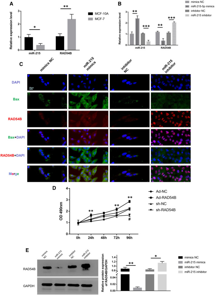
FIGURE 5.

(A) miR‐215 and RAD54B mRNA levels in MCF‐10A and MCF‐7 were determined by qRT‐PCR. U6 or GAPDH served as an internal control. (B) miR‐215 and RAD54B mRNA levels in MCF‐7 treated by mimics NC, miR‐215 mimics, inhibitor NC and miR‐215 inhibitor were determined by qRT‐PCR. U6 or GAPDH served as an internal control. (C) Immunofluorescence analysis of RAD54B and Bax in MCF‐7 cells treated by mimics NC, miR‐215 mimics, inhibitor NC and miR‐215 inhibitor. The nuclei were stained with DAPI. Scale bar = 20 μm. (D) Cell viability of MCF‐7 treated by Ad‐NC, Ad‐RAD54B, sh‐NC and sh‐RAD54B was monitored by the MTT assay. (E) Protein levels of RAD54B in MCF‐7 cells treated by mimics NC, miR‐215 mimics, inhibitor NC and miR‐215 inhibitor were determined by western blotting. GAPDH served as loading control. Data were representative images or were expressed as the mean ± SD of n = 3 experiments. *P < .05, **P < .01 and ***P < .001
