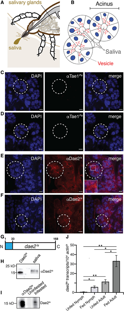
Figure 4. Dae2Is Is Found in Tick Saliva and Upregulated during Feeding.
(A) Schematic of the tick at the bite-site interface showing the salivary glands. The salivary glands produce the saliva that is injected into the host.
(B) Cartoon representation of salivary gland acini. Nuclei (blue), vesicles (red), and saliva flow routes (gray arrows) are shown.
(C–F) Confocal images of dissected I. scapularis salivary glands from adult females. Whole mounts were stained with DAPI (left panels, blue). Control mounts (C and D) were immunostained with rabbit antibody raised against a the bacterial Tae1Pa toxin (αTae1Pa). Experimental mounts (E and F) were immunostained with rabbit αDae2Is. Merged images are shown (right). Scale bars, 15 μm. Selected acini are marked (dashed white lines). A zoomed-in region is shown (box).
(G) Diagram of the dae2Is gene structure showing the predicted N-terminal signal sequence from nucleotide position 1–33 (blue).
(H) Western blot of collected saliva from partially fed I. scapularis adult females demonstrating presence of Dae2Is in saliva. First lane has recombinant Dae2Is as a positive control for rabbit αDae2Is reactivity.
(I) Western blot showing reactivity of mouse serum to Dae2Is. Recombinant Dae2Is protein was probed with mouse serum before (uninfested) or after I. scapularis feeding (infested). Rabbit αDae2Is was used as positive control for reactivity (first lane).
(J) dae2Is expression (normalized against actinIs transcripts) before (unfed) and after feeding (fed) on mice for nymph and adult ticks. *p < 0.05, **p < 0.01.
See also Figure S5.

