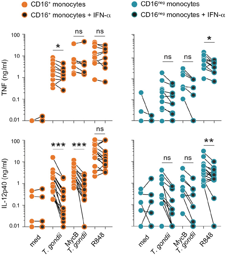
FIGURE 4: Pretreatment with type I IFN diminishes the IL-12 response of CD16+ monocytes and fails to enhance cytokine production by the CD16neg subset.
MACS-purified human primary CD16+ and CD16neg monocytes (106/ml) were cultured overnight in the presence or the absence of IFN-α (10 ng/ml) and then left untreated (med) or exposed to untreated or MycB T. gondii tachyzoites (MOI 1:1) or activated with R848 (300 ng/ml). TNF and IL-12p40 were measured by ELISA in culture supernatants collected after 18 hours of stimulation. The connected symbols represent the amount of cytokine in supernatants from monocytes from the same donor (n=6-12) cultured in parallel in the absence (orange and blue filled circles) or presence of IFN-α (black filled circles). * P < 0.05, **P < 0.01, ***P < 0.001.

