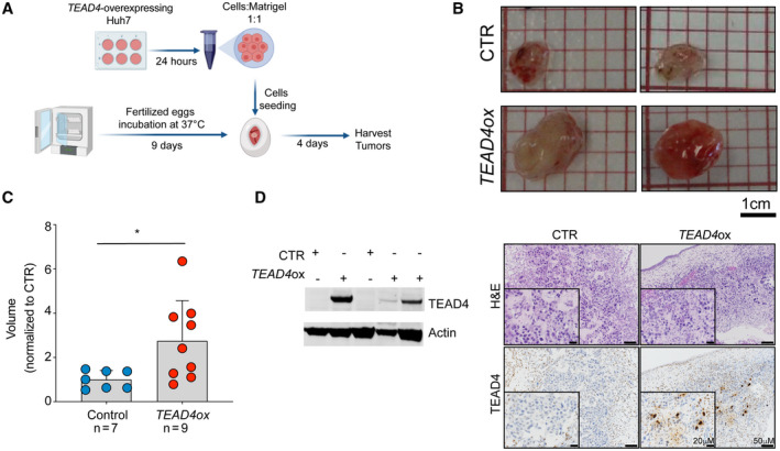
FIG. 2.

TEAD4 overexpression increases tumor growth in vivo. (A) Schematic representation of the CAM assay. (B) Photographs of TEAD4‐overexpressing or control Huh7 cells implanted in CAMs and grown for 4 days postimplantation. (C) Volume of tumors derived from the CAM experiment (n ≥ 7 tumors over two independent experiments). Values are normalized to the mean volume of control. (D) TEAD4 expression in Huh7 tumors extracted 4 days postimplantation analyzed by western blot (left) and IHC (right). Tumoral cells were immunostained with the TEAD4 antibody. Scale bars, (B) 1 cm and (D) 50 μm and 20 μm. Statistical significance was determined by the Mann‐Whitney U test; *P < 0.05. Data are mean ± SD. Abbreviations: H&E, hematoxylin and eosin; ox, overexpression.
