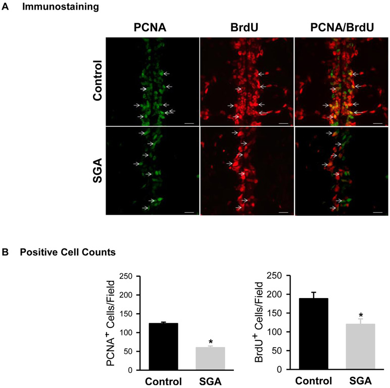
Figure 1: In Vivo Cell Proliferation.
Mid-line immunostaining of PCNA (green) and BrdU (red) incorporation in 1 day old Control (■) and SGA (■) males. (B) Quantification of PCNA and BrdU positive cells of 4 litters in each group and N=4 male pups per litter were studied. PCNA: t-value = 11.226, *P<0.001; BrdU: t-value = 3.208; *P<0.01 vs Control.

