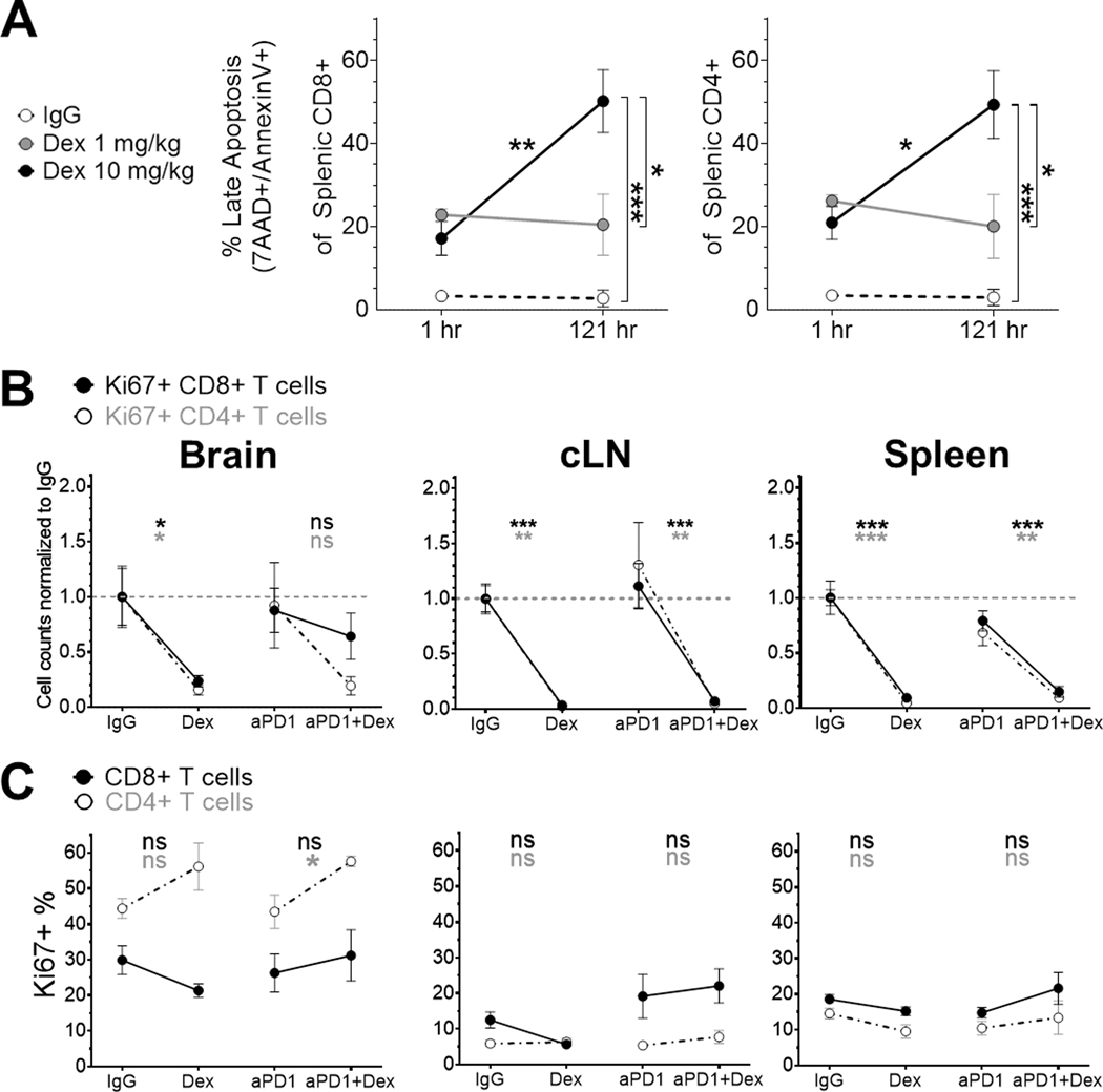
Figure 4. Concurrent dexamethasone increases apoptosis of CD4+ and CD8+ T cells in the GL261-luc2 glioblastoma mouse model.
(A) Late apoptosis was evaluated by 7-AAD+ and annexin-V+ staining in non-tumor-bearing mouse spleens (n=3/group, from a single experiment) either 1 hour after the first dexamethasone dose or 1 hour after the sixth daily dexamethasone dose. Apoptosis differences were tested by two-way ANOVA with post-test correction. Cell counts normalized to the corresponding IgG control group’s mean count (B) and percent (C) of proliferating CD4+ and CD8+ T cells were evaluated by Ki67 staining, using the same dosing schema and analyses as Figure 3 (n=4–8/group, derived from two experiments).
cLN, cervical lymph node; Dex, dexamethasone; hr, hour; ns, not significant, p≥0.05; *p<0.05; **p<0.01; ***p<0.001

