Figure 3.
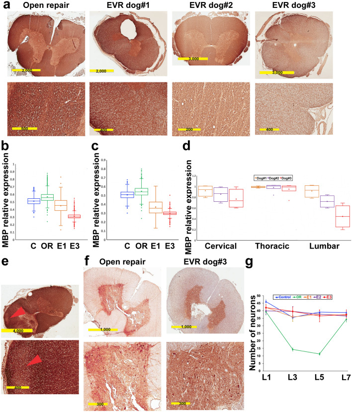
EVR primarily causes injury to the white matter. (a) Immunohistochemistry for myelin basic protein (MBP) in EVR dogs #1–3 versus OR. Cross-sections were at L5 level. (b), (c) Box and whisker plots showing MBP levels assessed by IHC on cross-sections of the SC throughout the L1–L5 region (b) or the T13–L3 region (c). C, control; OR, open repair; E1 and E3, EVR dog#1 and dog#3, respectively. Horizontal bars show the lower (first quartile), the median (second quartile) and the upper (third quartile) quartiles from bottom to top. The cross represents the mean value. (d) Box and whisker plots showing the dispersion of the mean levels of MBP expression in the cervical (C1, C3, C5 and C7), thoracic (T3, T5, T7, T9, T11 and T13) and lumbar (L1, L3, L5 and L7) regions of the SC of EVR dogs #1–3. (e) Reduced MBP level (arrowheads) in the cervical SC of EVR dog#2. (f) IHC for neurofilament heavy polypeptide in EVR dog#3 versus OR. Cross sections were at L5 level. (g) Neuronal density at the L1, L3, L5 and L7 levels in a control dog, an OR dog, and EVR dog#1 (E1), dog#2 (E2) and dog#3 (E3) was quantified by assessing the number of neurons per 10X field (given as mean ± standard deviation) as determined by pyruvate dehydrogenase testing. Scale bars are in µm.

