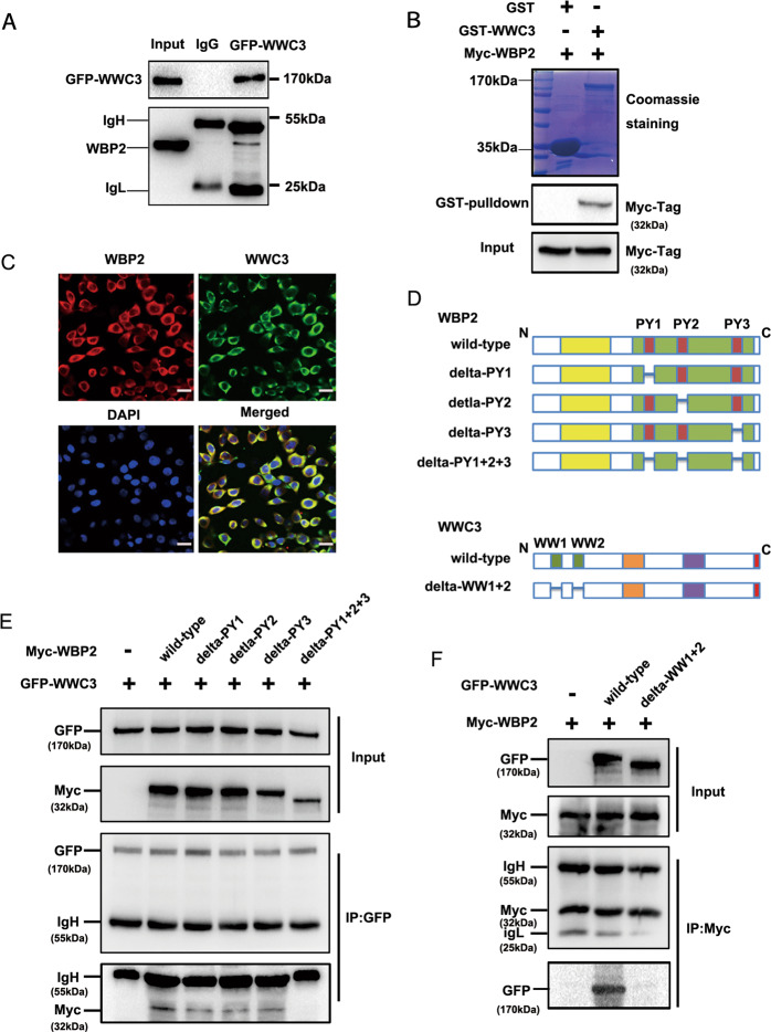
Fig. 5. WBP2 interacts with the WW domain of WWC3 via PPxY motifs.
A GFP-WWC3 was transfected into H1299 cells, and after 48 h, the cells were collected and lysed. A GFP monoclonal antibody was used for pulldown. The presence of WBP2 in the precipitate was detected by western blot analysis using a WBP2 antibody. B After incubation with purified GST or GST coupled-WWC3 protein for 6 h at 4° C, the binding status of two proteins was examined using Coomassie brilliant blue staining and western blot analysis. GST, glutathione-S-transferase. C Immunofluorescence assay results indicate that WBP2 and WWC3 colocalize within the cytoplasm of A549 cells. Magnification: ×400, scale bar: 50 μm. D Schematic diagram of WBP2 and WWC3 splicing mutants. E GFP-WWC3 and Myc-WBP2 wild-type or a series of mutants were co-transfected into H1299 cells. After 48 h, the cells were collected and lysed. GFP antibody was used for precipitation, and the presence of WBP2 was detected by immunoblotting using a Myc antibody. F Similarly, Myc-WBP2 and GFP-WWC3 wild-type or GFP-WWC3-△WW mutants were co-transfected into H1299 cells. GFP antibody was used for precipitation, and the presence of WBP2 was detected by immunoblotting using a Myc antibody.

