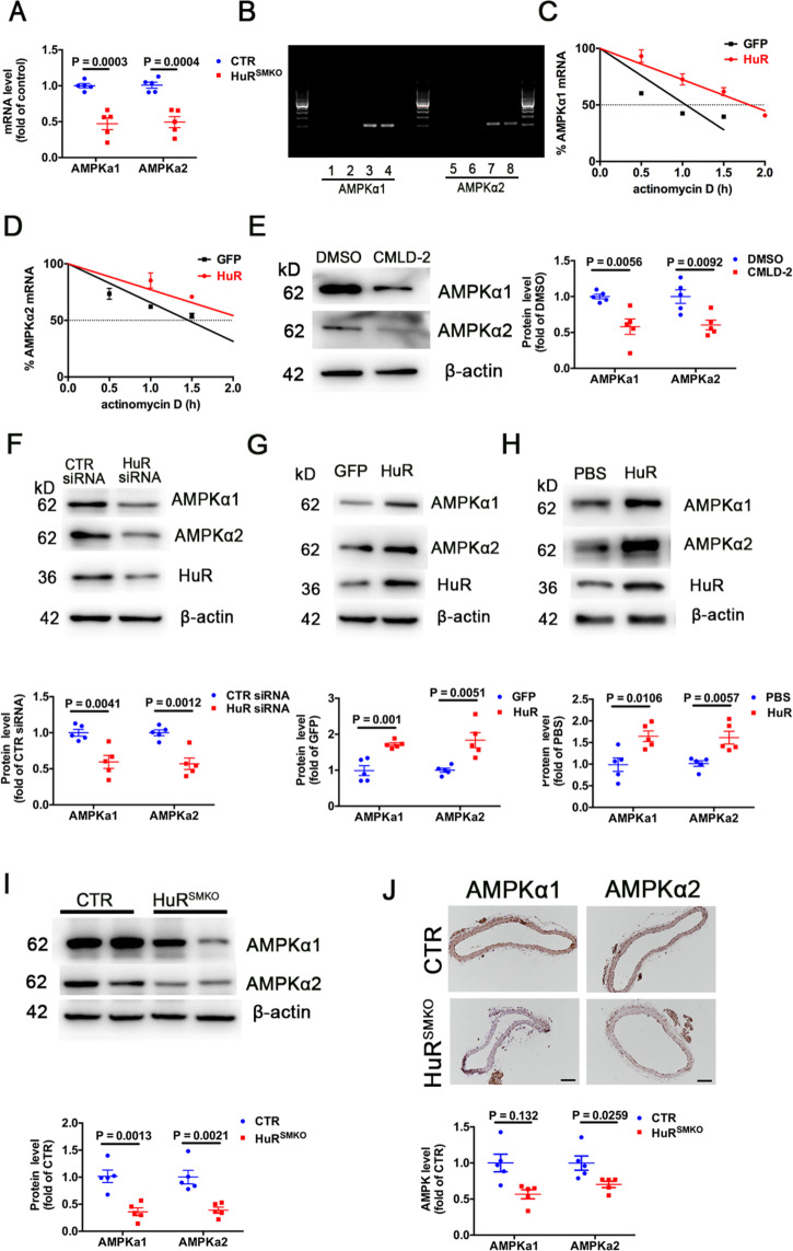
Fig. 5. AMPKα1 and AMPKα2 are the target genes of HuR.
A Quantitative RT-PCR analysis of aortic mRNA levels of AMPKα1, AMPKα2 from CTR and HuRSMKO mice (n = 5). B RNA immunoprecipitation with anti-HuR or control IgG antibody. Lanes 1, 5, no template PCR control; lanes 2, 6, IgG RNA immunoprecipitation; lanes 3, 7, anti-HuR RNA immunoprecipitation; lanes 4, 8, 10% input. C, D VSMCs were infected with adenovirus-expressing GFP (green fluorescent protein) or HuR and then treated with actinomycin D (5 μg/ml). Quantified RT-PCR analysis of percentage mRNA levels of AMPKα1 (C) and AMPKα2 (D) (n = 5) in VSMCs. E Western blot analysis of AMPKα1 and AMPKα2 in VSMCs treated with DMSO or 30 μM CMLD-2 for 24 h (n = 5). F Western blot analysis of AMPKα1 and AMPKα2 in VSMCs transfected with CTR siRNA or HuR siRNA for 48 h (n = 5). Western blot analysis of AMPKα1 and AMPKα2 in VSMCs, G infected with ad-GFP or HuR (n = 5), and H treated with PBS or 0.5 μg/μl recombinant HuR protein for 48 h (n = 5). I Western blot analysis of AMPKα1 and AMPKα2 in aortas from CTR and HuRSMKO mice (n = 5). J Immunohistochemical staining of AMPKα1 and AMPKα2 in aortas from CTR and HuRSMKO mice (n = 5). Scale bar = 100 μm.

