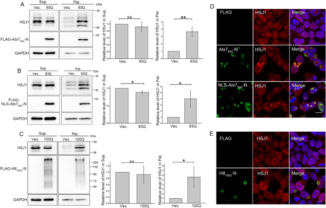
Figure 5.
PQE Atx7 and Htt sequester endogenous HSJ1 into aggregates. (A) Sequestration of endogenous HSJ1 by Atx793Q-N172. (B) As in (A), by NLS-Atx793Q-N172. (C) As in (A), by Htt100Q-N90. HEK 293T cells were transfected with each indicated plasmid and the lysates were subjected to supernatant/pellet fractionation and Western blotting. Indicated proteins were detected by using anti-FLAG, anti-HSJ1 and anti-GAPDH antibodies. The three main bands indicate different isoforms of endogenous HSJ1. Sup., supernatant; Pel., pellet. Data are shown as Means ± SEM (n = 3). *p < 0.05; **p < 0.01; N.S., no significance. (D) Immunofluorescence imaging showing that Atx793Q-N172 sequesters endogenous HSJ1 into inclusions. (E) As in (D), Htt100Q-N90. HEK 293T cells were transfected with indicated plasmids and then subjected to immunofluorescence imaging. PolyQ proteins were stained with anti-FLAG antibody (green), HSJ1 was stained with anti-HSJ1 antibody (red), and nuclei were stained with Hoechst (blue). Scale bar = 10 μm.

