Figure 7.
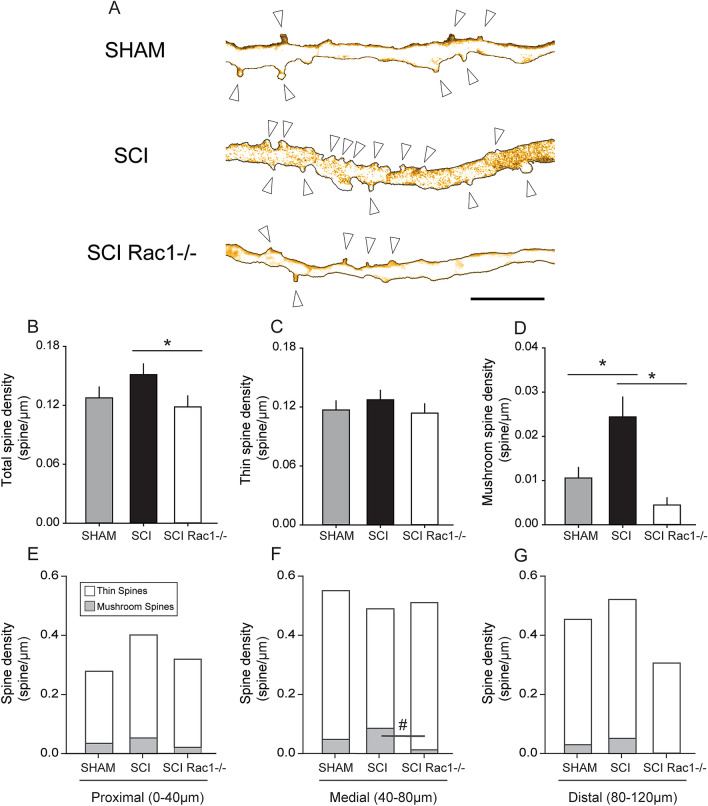
Conditional Rac1 KO in alpha-motor neurons reduces abnormal dendritic spine morphology associated with SCI and hyperreflexia. Analysis of dendritic spine profiles reveals differences in (B–D) spine density and (E–F) distribution. (A) Reconstructed dendritic segments from tdTomato filled spinal motor neurons showing apparent differences in dendritic spine profile between Sham, SCI and SCI Rac1−/− (arrow denotes spine). (B) Total spine density, which includes all spines, was significantly lower in the SCI Rac1−/− compared to control (* = p < 0.05). (C) There was no difference in the density of thin-shaped spines between groups. (D) SCI induced a significant increase in mushroom spine density compared to Sham (* = p < 0.05). In contrast, SCI Rac1−/− reduced mushroom spine density compared to SCI (* = p < 0.05). (E–G) Assessment of dendritic spine density within the (E) proximal (0–40 µm), (F) medial (40–80 µm) and (G) distal (80–120 µm) dendritic branches of tdTomato filled motor neurons. (E, G) Dendritic spine density was not different within the proximal or distal region between Sham, SCI and SCI Rac1−/−. (F) SCI induced an increase in mushroom spine density in the medial region, which was not observed in SCI Rac1−/− animals (# = p < 0.05). Graphs are mean ± SEM. Scale bar in (A) = 10 µm.

