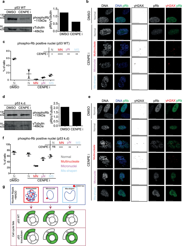
Fig. 5. Multinucleated cells uniquely display reduced phospho-Rb despite compromised p53.
RPE1 p53 WT (a–c) or RPE1 H2B-GFP p53 k.d. d–f cells were treated with DMSO or CENPEi for 16 h and 48 h later cells were immunostained with antibodies against pRb and gamma H2AX, or cells were lysed for immunoblot. a & d Immunoblot shows pRb or gamma-tubulin levels in RPE1 p53 WT (a) or p53 kd (d) cells following DMSO or CENPEi treatment, as indicated. Note the gamma-tubulin is the same as displayed in Supplementary Fig. 1. Right panel shows a graph of pRb fluorescent intensity, normalised to gamma-tubulin. Representative images of nuclear atypia following CENPEi treatment of RPE1 p53 WT (b) or p53 kd (e) cells. Scale 15 μm. Graph of proportion of pRb positive or negative RPE1 WT (c) or p53 kd (f) cells, after DMSO or CENPEi treatment. N > 100 for WT or >150 for p53 k.d cells from 2 or 3 independent repeats (shown as shades of grey), respectively. Statistical analysis using multiple unpaired t-tests, comparing each morphology after CENPEi treatment to normal nuclei after DMSO. g Model comparing nuclear atypia shows large-scale DNA damage in multinucleate cells, but not in misshapen nuclei, and in micronucleated cells, gamma H2AX foci are majority confined to the micronucleus. Nuclear atypia causes G0 arrest in p53 WT. In p53 k.d. conditions, only multinucleate cells are G0 arrested.

