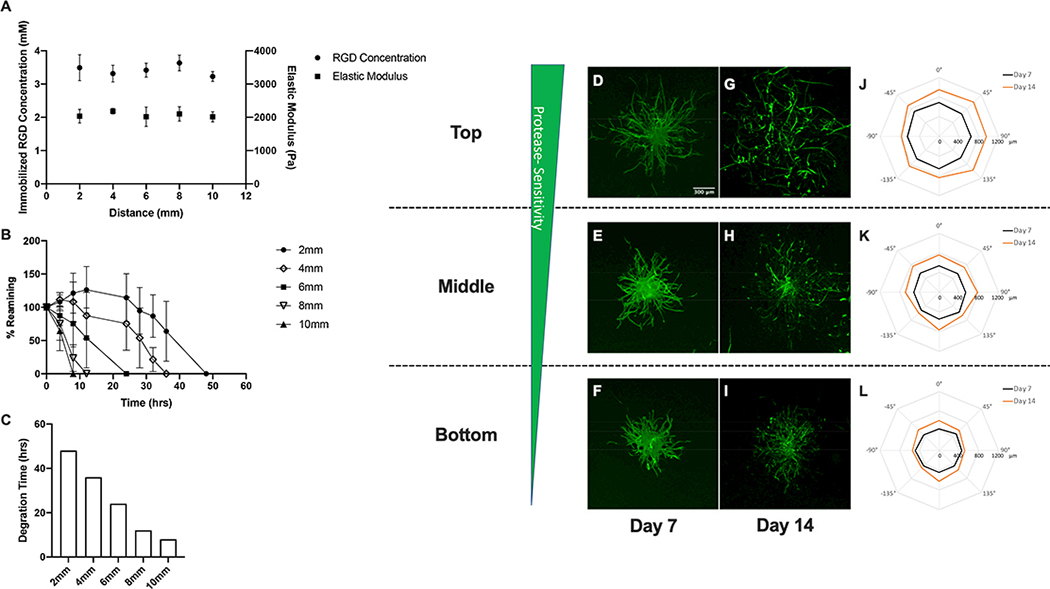
Figure 5:
Characterization of spatial variations in matrix properties and vascular sprout formation in protease-sensitivity gradient hydrogel scaffolds. (A) Quantification of spatial variations in immobilized RGD concentration and elastic modulus, (B) degradation kinetics and (C) time required for complete degradation in collagenase enzyme solution. Confocal images of phalloidin-stained spheroids embedded within top, middle and bottom regions of hydrogel scaffolds with gradients in elastic modulus after 7 and 14 days culture (D-I). Polar plots of maximum sprout length by angle of spheroids encapsulated in different regions along the gradient (J-L).

