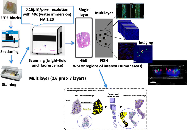
Fig. 1.
Workflow of tissue sectioning, staining and scanning. Serial sectioning of FFPE tissue blocks was used for H&E or IHC staining in order to characterize the region of interest. H&E and IHC slides were scanned at wide-field mode with 20× water immersion objective at a single layer. ROIs on FISH slides were scanned at confocal mode with multiple layers (N = 7 layers at 0.6 μm interval) with 40× water immersion objective and a final image resolution of 0.16 μm/pixel. Three filters were chosen: DAPI (blue), FITC (green) and TRITC (red). Showing in a dashed line frame: we are currently developing a deep learning algorithm for an automated tumor area detection

